|
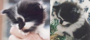
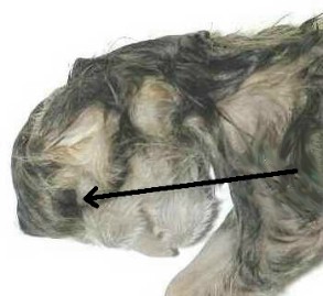
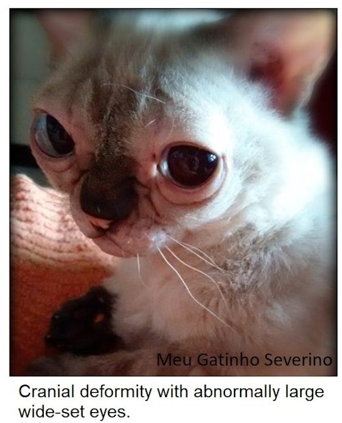
The peculiar shaped face of this five week old black-and-white kitten is due to hydropcephaly and anophthalmia. |
|
|
FELINE MEDICAL CURIOSITIES: FACIAL DEFORMITIES
Hydrocephaly, Anencephaly etc
Note: Contrary to suggestions on some bulletin boards, the images here are not photoshop. With the exception of those labelled as artist's impressions these are photos of medical conditions. These pages are intended as a medical reference site. Offsite links to images on these pages is not supported - bandwidth costs money!
HYDROCEPHALY
|
The peculiar shaped face of this five week old black-and-white kitten is due to hydropcephaly and anophthalmia. |
|
|
Hydrocephaly means fluid accumulating inside the skull. It causes a distinctive domed appearance. While the kitten is young, the skull grows increasingly domed or bulbous to accommodate the fluid. The tall forehead is characteristic of the condition. Later, the skull cannot stretch and the build-up of fluid causes pressure on the brain, increasing brain damage and eventually death. Nicknamed "the dog-faced kitten", the black-and-white kitten (above) was put to sleep at about 8 weeks old because of increasing fluid pressure and poor prognosis. In hydrocephalic humans, shunts are used to drain the fluid; at present shunts are not available for cats with the condition.
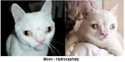
Some case of hydrocephaly do survive. "Moon" was a hydrocephalic kitten with hare lip and cleft palate and is owned by Leila in Sao Paulo, Brazil, who sent these photos. According to Leila, Moon was found abandoned at 4 months old. He has undergone surgery to repair his hare lip and cleft palate and, at the time of writing, was a year old and doing well. I believe this to be hypertelorism (wide face) rather than hydrocephaly as the head is wide, rather than domed.
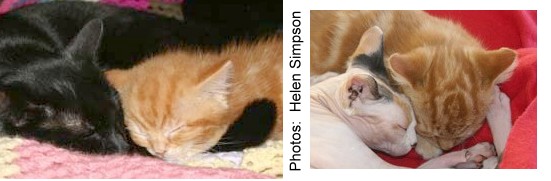
Helen Simpson (Cambridgeshire, UK) provided these photos and details of her hydrocephalic kitten, Josh. Josh's case was not too severe and he has survived kittenhood and is doing well. When Helen got Josh from a rescue in Kent, he he was due to be put to sleep because of the shape of his head and the organisation he was with said no-one would want him. Luckily someone told Helen about Josh's case. His head is quite domed and to begin with the vets were not sure if he would live because affected kittens rarely survive as their mothers reject them or they have far more severe cases of hydrocephaly. As he's grown older he has "grown into" his head (it is less out of proportion) although it is still very domed. As a small kitten, his head resembled a balloon on his tiny body.
|
|
Josh was a strange kitten with "no real sense of reason but bags of personality." To begin he often ran into things and when running, his brakes didn't work. When working out how to do things, for example how to jump on a bed, he squeaks and chatters to himself about it. He was 8-9 months old in April 2007 and has been neutered (using a different anaesthetic from normal due to his condition).
MENINGOENCEPHALOCELE
(Incomplete skull with protrusion of meninges and brain)
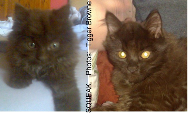
Squeak (owned by Tigger Browne) was a rescue kitten with a cranial abnormality. By 4 months her skull did not cover her brain properly and her face is twisted from just above her nose. Leading from the top of her nose is a very big V-type groove which leads to a soft lump on the top of her head. The soft lump on her head is a meningoencephalocele - the protrusion of her brain through the gap in her skull. Usually kittens with meningoencephalocele are stillborn, die very early or are put to sleep, so Squeak may be unique in having survived to 4 months. A skull x-ray would determine how much skull is missing and how much brain is protruding through the gap. In addition, Squeak's gender is ambiguous and s/he has a small umbilical hernia (Squeak will be referred to as "she" for convenience).
|
|
Because the brain had developed and was growing abnormally resulted in seizures and abnormal behaviour: panic attacks involving foaming at the mouth and wetting herself and showing signs of terror. Seizures lasted for 2 - 3 minutes after which she became very clingy and loving and tried to clean herself obsessively (she had problems cleaning her head and her rear end). In general, her behaviour was scatty, but not intellectually retarded. She appeared to hallucinate, morseso that cats that normally chase invisible prey. Her movement tended to swing when she walked and she frequently missed her target when jumping or pouncing; her owner believed Squeak may have had some impairment of vision or spatial awareness. The seizures were are likely to get worse, eventually leading to ataxia (lack of co-ordination), possibly blindness and probably increasing paralysis. The brain is also very easily damaged where it protrudes; any trauma could cause swelling. It's not possible to give a prognosis in months or years because Squeak is unusual in surviving this far. In some cats, this is inherited (American Shorthair Craniofacial Defect, Burmese Head Defect), though it can occur at random through mutation or developmental abnormality.
Apart from the seizures and strange behaviour, Squeak weaned quickly and ate and drank normally and was quick to learn to use the litter tray. She was fully housetrained except when having a seizure. She was underweight when adopted, but grew to a healthy 2 kg (4.4 lbs) and was a lively, happy, inquisitive little soul that loved playing with feet or running around like any other kitten causing mayhem wherever she went. Tigger is experienced with epileptic animals and rescue cats and monitored Squeak's condition. It wasn't known whether anti-epileptics would control the seizures. As Squeak (still sexually ambiguous, but possibly developing as a male) grew her fits initially decreased in frequency and intensity. She went through the normal shedding of kitten teeth and the dome-shaped swelling on Squeak's head became less pronounced, especially on the right side, as she grew. She also became able to clean herself. Unfortunately on Saturday 13th December 2008, Squeak became hyperactive and had 3 increasingly violent fits in succession. As her fits were becoming continuous and more distressing due to the pressure on the brain, she was put to sleep aged approximately 5 months old. Squeak appears to be the oldest recorded survivor of meningoencephalocele.
UNEVEN FACIAL DEVELOPMENT

Sarah Saylor provided these photos of a 5 and a half week old kitten "Batty Cat" with facial deformites. It appears that one side of Batty's face hasn't developed as fully as the other side - giving him a smaller eye and twisting his nose and mouth to one side. As long as he can eat and drink normally and the rest of his body is unaffected by developmental deformities, Batty should have a normal life. The cleft lip is cosmetic and there is no cleft palate; the lip could be repaired surgically if it caused any problems. The twisting of the nose could cause noisy or obstructed breathing, but that could be helped by surgery. One eye is under-developed and appears to have limited sight. As Batty grows, a vet will need to check his jaw alignment (it may grow unevenly) in case there's any twisting or under-development on one side in which case he might need some adult teeth removed to avoid over-crowding. Batty was otherwise in good health and had a good appetite, though it has been a little harder for him to move onto soft food due. He was bottle-fed from birth.

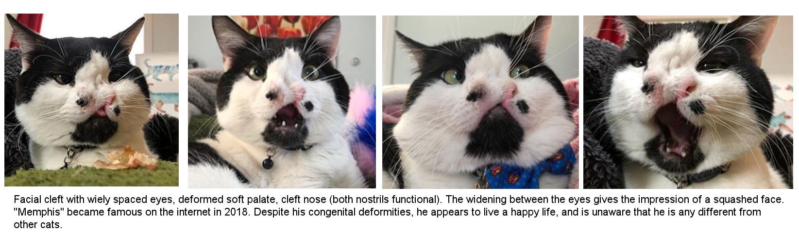
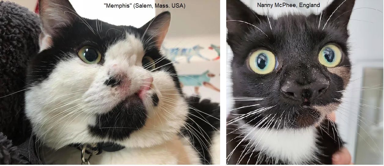
In 2018, a black-and-white cat called Memphis was taken to a rescue group in Salem, Massachusetts. Other shelters threatened to euthanize Memphis when his owners had to give him up. Memphis had been born with two functional noses (more precisely wide-spaced nostrils with fur between), a deformed soft palate and cleft lip, and facial congenital abnormalities. In 2023, a similar cat, also black-and-white, called Nanny McPhee, was taken to a shelter in Warrington, England.
This type of deformity - an abnormally wide distance between the nostrils - is often associated with cleft palate and cleft lip. There is a long-snouted South American breed of dog (Double-Nosed Andean Tiger Hound) which has 2 wide-separated nostrils rather than a fused nose, but no other face or mouth abnormalities. Neither the double-nosed dog (where the trait is genetically inherited), nor the cats (where it is a birth defect) have an enhanced sense of smell.
FACIAL DERMOIDS
A dermoid cyst is a type of teratoma that frequently consists of skin, hair follicles, and sweat glands (larger cysts can contain teeth or other mature tissue). Visually they appear as abnormally located patches of skin or whisker pad. In cats, dermoid cysts are associated with the Burmese and American Shorthair craniofacial defect where cats were bred for domed heads. They can also occur randomly, either due to a developmental anomaly in the embryo or as part of a genetic condition where there may also be asymmetrical facial development, crooked nose or jaw, cleft lip, severe cleft palate or duplicated or wrongly-placed canine teeth.
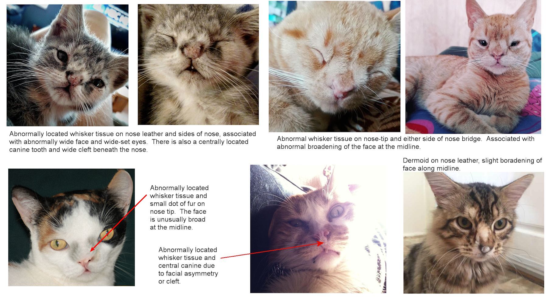
They can occur when skin cells and things like hair, sweat glands, oil glands or fatty tissue get trapped in the skin as they embryo develops. In many of the cases shown here, the face is abnormally broad at the midline which shows that the cysts are linked to other issues and not simply to trapped skin cells. Extreme broadening can lead to duplication of facial structures e.g. eyes, nose bridge, but a single lower jaw.
TERATOMA ON FOREHEAD OF PUMA CONCOLOR
Deformities also occur in big cats. A mountain lion shot in southeast Idaho, in 2015, had a partial jawbone and set of teeth growing out of its forehead. In December 2015, Tyler Olson reported that a mountain lion had attacked his dog at his rural Weston Canyon home. A group of mountain lion hunters and neighbours quickly tracked the cougar through Weston Canyon (about 8 miles southwest of Preston near the Idaho/Utah border) and shot it (this is legal). The hunter reported the animal's deformity to the authorities when reporting the kill. A conservation officer inspected the kill and sent photos of the lion s deformity to Idaho Fish and Game s Southeast Regional Office in Pocatello. The year-old lion had a partial jawbone and fully-formed teeth, including what appeared to be small whiskers, growing out of hard, fur-covered tissue on the left side of its forehead, an abnormality that local Fish and Game biologists had never seen before.
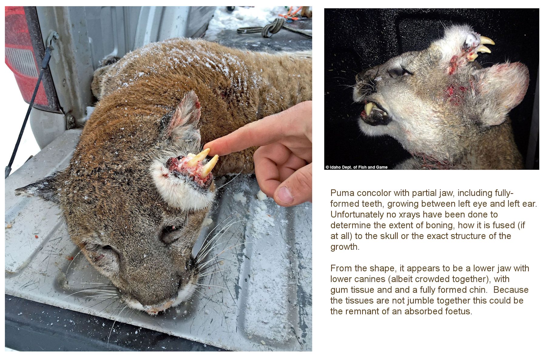
Experts have not been able to agree on the cause of the unusual mutation. It could be the remnants of a conjoined twin which died in the womb, or incomplete duplication of facial features; the fact that the tissues are well organised into a lower jaw tends to support this. They feel that a more likely explanation is that it is a teratoma, a type of tumour that can grow other body tissues such as hair, teeth, bones - even eyes. Though extremely unusual, there are well-documented cases of teratomas in humans, dogs and horses, however the tissues are often disorganised. Less likely is that the mountain lion suffered a jaw injury that healed in an abnormal. There is no evidence of an injury to the mouth, and it appears to have a normal set of teeth in its mouth. Idaho Fish and Game's Southeast Regional Office, in Pocatello, had hoped to bring the carcass in for examination. X-rays and analysis could have revealed the cause of the abnormal growth. Officials asked the hunter to bring the cougar in for testing. However, the hunter never brought the animal in and is not required to do so by state law. According to rumour, the cougar is now a taxidermy specimen in somebody s home.
MISCELLANEOUS CRANIAL AND CRANIOFACIAL DEFECTS
Cranial refers to head and skull defects that affect the braincase. Craniofacial defects affect the head and face: the soft tissues and the bones forming the front of the skull i.e. the face and jaws. These abnormalities are often related to other conditions such as dwarfism, hydrocephaly or metabolic disease. These include extreme over-bite/under-bite, hare lip (cleft lip) and cleft palate.
Craniofacial anomalies are an intrinsic part of some breeds such as the modern ultra-typed Persian and the now-extinct Peke-Face Red Persian (extreme brachycephaly) and the extremely elongated head of the modern Siamese. The Peke-Face Persian is different from the modern "piggy" Persian and the last known true Peke-face Persians was registered in 2002, and only about 100 such cats were ever registered. Their skull structure differed greatly from the standard Persian: a very round head with a very strong chin and very wide-set eyes. The muzzle was wrinkled and the nose pushed inward and indented between the eyes, plus there there was an additional horizontal break located between the usual nose break and the top dome of the head. This second break created half-moon boning above the eyes (brow ridge and double dome) and an additional horizontal indentation (dimple) in the center of the forehead. These features were part of the breed standard and caused a lot of problems. Kittens tended to have heads too large for them to be born without a caesarian section. Their palates could be too high so that they couldn't suckle. Their tear ducts were malformed and did not drain (a problem seen in extreme Persians today). In the wild, the kittens and their mother would not have survived, so this was a deferred lethal mutation. Probably multiple genes were involved rather than a single gene, and these genes were combined together because of inbreeding.
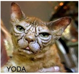
Inbreeding is a problem in cats with small gene pools. Yoda, below, has thickened facial skin giving a puffy appearance. The thickened facial skin does not have working hair follicles, so Yoda lacks fur on his face and lacks whiskers.
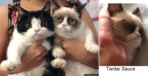
A dwarf cat named Tardar Sauce (born April 4, 2012), Tabatha Bundesen, became an internet meme in the guise of Grumpy because her condition gives her a flat face, protruding eyes, an under-bite and a grumpy facial expression. Tardar Sauce (a colourpoint with white) and her brother (black-and-white bicolour), also a dwarf, called Pokey were born to normal parents. As well as their distinctive faces, they are undersized and have limb deformities associated with feline dwarfism.
Tabby and white Lil Bub (2013), owned by owner Michael Bridavsky, had a serious and rare bone condition that caused her paws to be deformed and her tongue permanently protruding due to a very short lower jaw. Lil Bub's face had a distinct "stop" (change of angle) between the wide set eyes, an extremely under-developed mandible (lower jaw) and she lacked teeth. She was also a polydactyl with six toes on her paws. She stopped growing at 7 months old and weighed only 1 kg throughout her life. She died in 2019, aged 8 years old, still looking like a kitten.
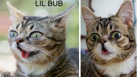
Daniel Ibrahim and Dario Lupanez in Berlin requested permission to investigate Lil Bub s genome. This would help gain information about similar human diseases. They identified the mutations responsible. One mutation was in the Sonic Hedgehog Gene which is responsible for the formation of the digits and which causes polydactyly in vertebrates. Another mutation affected the RANK gene (TNFRSF11A) responsible for osteoclast function. A mutation in the RANK gene is associated with osteopetrosis ( stone bone disease ) in humans and mice, and caused a similar state in Lil Bub. It was because of this mutation that she never grew teeth because her jaw bones hardened too quickly and didn't allow the teeth to erupt from the bone.
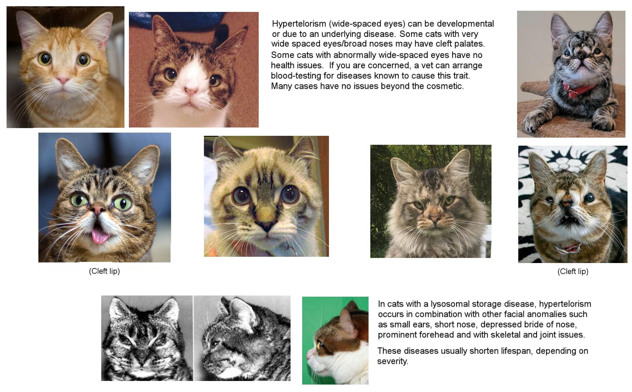
HYPERTELORISM (WIDE SPACED EYES) & OTHER CRANIAL ANOMALIES
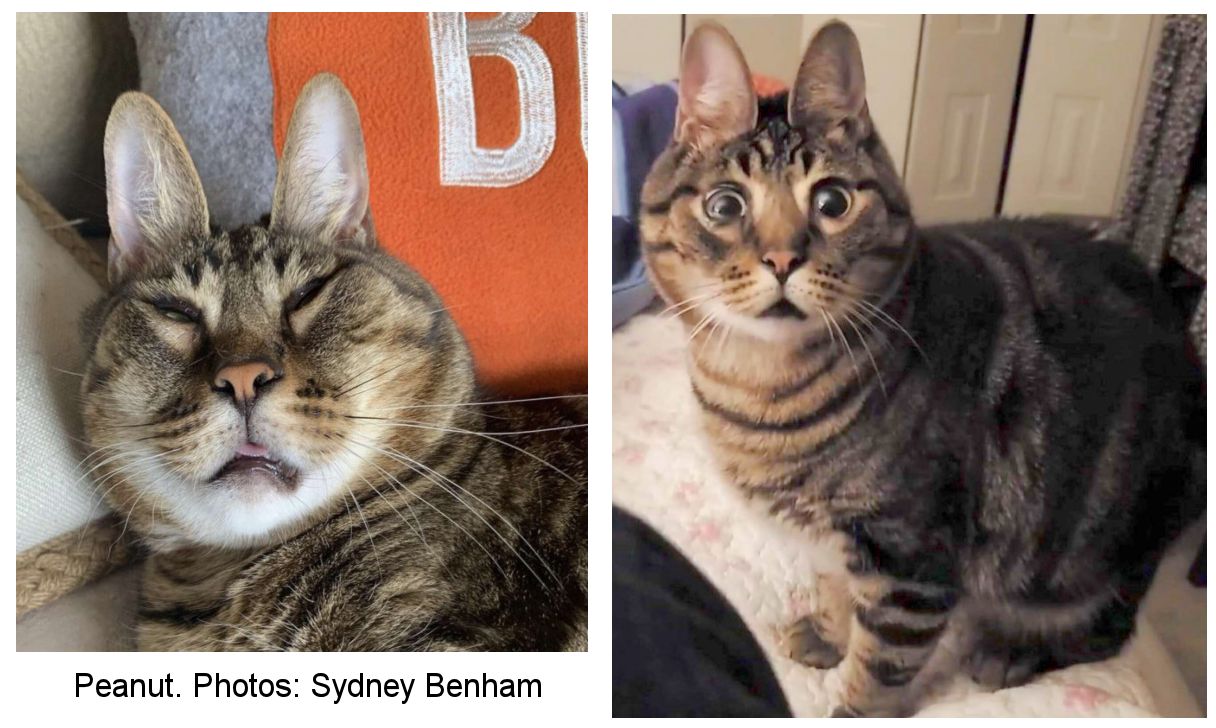
Peanut, aged 8, and nine other cats went to a rescue shelter when their owner died. Peanut is deaf and partially blind, and has a facial deformity that makes him resemble a rabbit. He was adopted by Sydney Benham in 2020. Despite his disabilities, he is a loving and happy cat with a good quality of life. The cause of his unique appearance is not know, but is likely a random mutation.
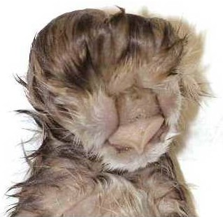
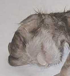
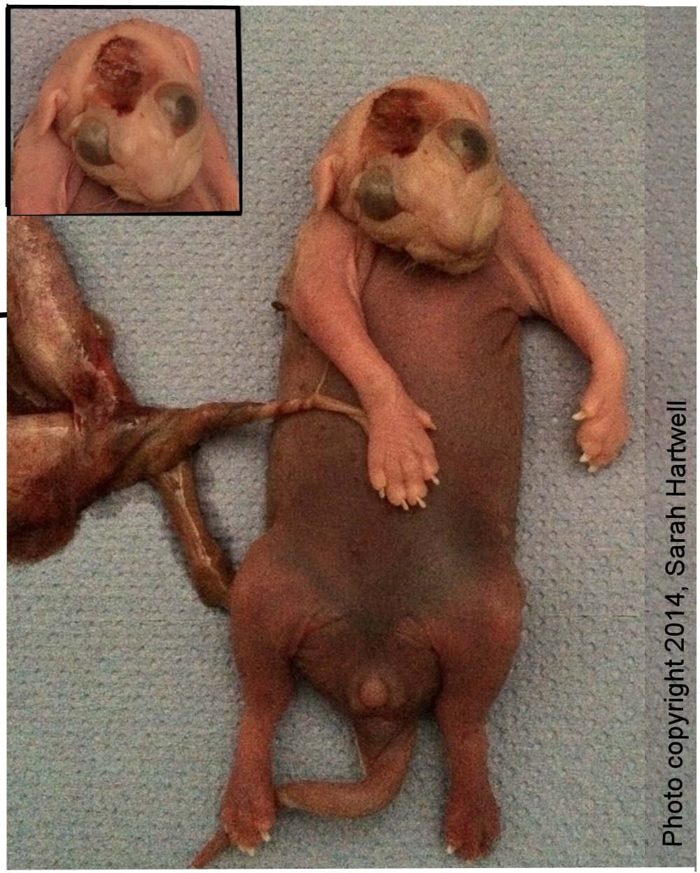
THE AMERICAN BURMESE & AMERICAL SHORTHAIR CRANIOFACIAL DEFECTS
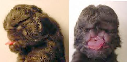
Some American lines of Burmese cats are genetically prone to head abnormalities due to breeding for a domed skull (European Burmese are more oriental in shape and apparently free of this defect). As seen above, the upper part of the muzzle and roof of mouth are duplicated and the area above the muzzle is incomplete. There is only one lower jaw and tongue. The ears and eyes are malformed. The skull doesn't close completely, leaving a protruding area of brain covered only with skin and not protected by bone. It is necessary to humanely destroy any kittens that don't die at birth. Because such deformities invariably involve brain damage, it is merciful that the kitten died within moments of birth.
The Burmese craniofacial defect, or median cleft syndrome, is scientifically known as Frontonasal Dysplasia (FND3) and is associated with a mutation of Aristaless-Like Homeobox 1 (ALX1). Studies demonstrated that it is a simple co-dominant trait i.e. it inherited recessively; carriers have a desirable (in breed-defining terms) modified head shape, but homozygotes are lethally affected. A mutated ALX1 gene is present in affected Contemporary Burmese and absent from unrelated cats with brachycephalic features.
the late 1970 s, a male American Burmese cat with a more brachycephalic head type, that was inherited by his offspring, became a highly popular sire, resulting in the Contemporary Burmese look. When Contemporary Burmese were bred together, a craniofacial defect appeared in 25% of offspring. It was inherited as an autosomal recessive, but carriers of the mutation are more brachycephalic than non-carriers and were positively selected for in the breed. Because carriers are visually identifiable, the trait is also described as co-dominant (or as dominant with additive effect). Affected kittens were generally born live, but had to be euthanized because the condition is incompatible with life. The heterozygous (gene carrier) cats became the standard phenotype of the breed and the predominant winners at cat shows.
Traditional lines of Burmese are less extreme, but have been selected over 3 decades for a more rounded head type that is not associated with congenital abnormalities. Hence some Traditional lines became difficult to distinguish phenotypically from the affected contemporary lines. The Burmese is also one of the most genetically inbred cat populations worldwide and suffers several other health concerns. The craniofacial defect led to American Burmese lines being banned by many other registries in order to keep them free of the head defect. This meant a gene test was needed in order to eliminate the ALX1 mutation from the American Burmese.
The mutation was also found in the American Bombay (developed from the American Burmese x American Shorthair) and also in the American Shorthair (where Burmese x American Shorthair hybrids entered the American Shorthair gene pool), but not in the Australian Mist, Burmilla, European Bombay or Asian as these were developed from non-American Burmese. It also wasn t found in the Tonkinese as these were not selected for an extreme head type hence the gene carriers were selected against.
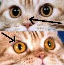
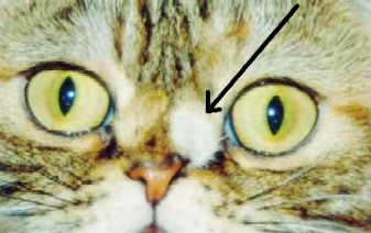
Similar defects appeared in the American Shorthair breed since the 1970s/1980s and may also affect the American Wirehair. The most minor forms of the defect were overlooked as "merely cosmetic" by breeders as the cats generally had superior conformation (wide spaced large eyes, short broad muzzle, concave profile, large boning) and were successful on the showbench. Show success made these desirable breeding animals and caused the spread of the defective gene. The "cosmetic" effects were actually indicators of the more serious defects that would show up when carriers were bred to each other. About 90% of cats with only 1 copy of the mutant gene had a "dot" (longitudinal depression) on their nose that was evident from birth. Others had dermoids (abnormally located patches of skin) and a few had more serious defects such as harelip, severe cleft palate, duplicated canine teeth, duplication of tongue tissue, crooked jaw, coloboma (fissure of the eye) through to lethal athymia (missing or non-functional thymus gland) or seizures (indicating brain abnormalities).
The dermoids were usually pieces of whisker pad, complete with small whiskers, growing in abnormal places on the face e.g. on the side of the nose, below the eye or even on the eye itself. Small dermoids were hard to spot e.g. a whiskery "mole" under a nostril. Breeders often snipped off tiny dermoids, believing them to be cosmetic and not realising they were symptoms of the head defect gene. The nose was often skewed and the jaw crooked, showing that the head had not formed symmetrically. Coloboma (fissure of the eye socket) is caused by the eyelid plate not developing properly; it looked as though the rim of the eye socket (brow) is crooked. These supposedly cosmetic traits were clear indicators that the cat carried the head defect trait.
When a cat with a "dot" on the nose was bred to unaffected cats, about 50% of the offspring inherited the "dot" and about 5% of the offspring inherited lethal defects. The 50% affected offspring also inherited one or more of the defects such as dermoids or harelip . This suggested the gene was an autosomal (not sex-linked) dominant. When 2 cats with dots on the nose were bred together, many more offspring inherited 2 copies of the gene, resulting in gross and lethal head deformities.
|
(a) "dot" on midline of nose |
(c) cheek-pad tissue growing on side of nose, distorting nose shape |
||
|
|
|
|
|
Kittens that inherited 2 copies of the gene were often born alive, but severe head defects meant they survived only a short time. The brain might be covered by skin but the bony skullcap was missing. The skin might cover a fluid filled chamber (hydrocephaly). The brain was grossly abnormal with little or no normal brain tissue; it ranged from holoprosencephaly (malformed forebrain) to anencephaly (no forebrain). Holoprosencephaly is a failure of the forebrain to divide into hemispheres or lobes and is accompanied by under-development of facial features such as the nose, lips, palate and teeth. In milder cases holoprosencephaly causes wide spaced eyes, harelips and cleft palate, but in severe cases it causes cyclopia (the eyes fuse into a single deformed central eye, often with a proboscis above it). Other affected kittens had small rudimentary eyes located on the sides of the head rather than on the face. Some had severe cleft palate, extreme harelip on both sides of the mouth, brachygnathia (short or receding jaw, flat face) and protruding tongue.
Because cats with minor defects such as the nose dot were often superior in other ways, breeders were unknowingly selectively breeding the cranial defect into the breed. The desire to "ultra-type" cats meant that breeders working with extreme types were more likely to breed affected cats together and encounter the head defect than breeders working with the more moderate or traditional types. Some of the affected cats had won high accolades on the showbench and were desirable breeding stock. The gross deformities would only show up a few generations later when cats with the mutation were bred to each other and kittens with lethal head defects were born.
The incidence of cleft palate in Persian cats (also noted for their wide set eyes, short muzzle and domed head) may also be related to breeding cats for domed heads (in Persians 2 genes are implicated in brachycephaly: CHL1 and CNTN6, which are known to determine face shape modification in humans).RAINE SYNDROME (UNCONFIRMED)
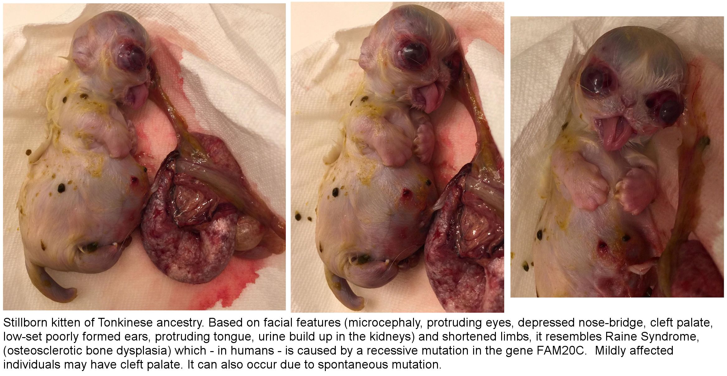
Below is a stillborn kitten whose features resemble Raine Syndrome. Milder forms of the syndrome include cleft palate. Severe forms are incomaptible with life. The bones harden abnormally early hence the small misshapen skull, with protruding eyeballs, and short limbs. The swollen belly may be due to a build-up of urine in the abdomen where the kidneys do not function properly.
CONJOINED KITTENS
For othe types of conjoining see Conjoined Kittens
ANENCEPHALY (ABSENCE OF FOREBRAIN)
The following information and accompanying images have been provided by Joen Seddon In December 2004 for the benefit of other breeders encountering gross deformities in kittens. Joen's tortoiseshell queen produced a litter of 4 kitten following a difficult labour. The first kitten arrived after approximately 3-4 hours after bearing down. The queen had lost a great deal of fluid (more than usual) before the first kitten arrived. The kitten was born tail first with the sac already broken. On first sight, the kitten was found to have extremely bulging eyes and no eyelids. Closer inspection indicated that there may have been eyelids (or skin) present, but they were completely transparent and the eye ball was completely visible. The pupils were large. The tongue was enlarged and everything from the bridge of the nose upwards appeared to be collapsed or absent. The kitten lacked a forehead and fontanel. The ears were approximately at nose level and facing outward rather than upward. On the top of the head was a small opening revealing a membrane. The kitten appeared very strong, but showed no interest in feeding.
After the remaining kittens were born, Joen removed the deformed kitten (and was surprised that it was still alive) to see if it had any instinct for food in spite of apparent lack of the forebrain. It had a good deal of strength, but lacked the co-ordination to initiate feeding, however it took kitten formula from a small dropper. Though it became excited at being dropper fed, it showed no sucking ability. When placed directly at the mother's nipple, it showed interest in feeding, but could not grasp the nipple and was only able to lick at it with its enlarged tongue. The inside of the mouth was badly deformed. The rest of the body appeared entirely normal and the weight was 200 grammes. At 9 hours, it was active and looked "like a bat". Joen decided to let nature take its course. The kitten survived only 20 hours. After 12 hours it began crying loudly, probably due to inability to feed (because it lacked a forebrain, it lacked the ability to feel pain; the crying was a reflex and did not indicate suffering (this has been studied in human cases of anencephaly)).
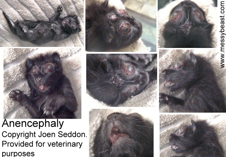
The mother cat did not treat the deformed kitten any differently from the three normal siblings. The surviving kittens are described as "normal, hefty and spunky" and also 200 grammes (approx). Oddly, the anencephalic ("lacking forebrain") kitten was also spunky, though the absence of the cerebrum meant the kitten lacked consciousness (feeding reflexes and breathing are due to the more primitive parts of the brain). Joen's photos were taken shortly after the kitten died and there are signs of dehydration. The eyes were even more pronounced in the hours after birth.
The mother had difficulty birthing the deformed kitten, but the remaining 3 kittens were birthed more easily. The mother had previously only been bred 3 times (all planned) with the same male and had produced a single kitten on each of the previous occasions. The latest 4 kittens had been conceived while her previous singleton kitten was only 3 1/2 weeks old and had been sired by a stray tom when the female escaped from the house. It was not realised that the queen was pregnant for a month. The cause of anencephaly is unknown, but human cases are linked to the mother's diet and vitamins, particularly to folic acid deficiency. Gross neural tube deformities due to genetic causes have been reported in the Manx breed, but the kittens are usually reabsorbed early in gestation or arrive as partially reabsorbed ("mummified") kittens. It is therefore possible that the previous singleton litters were due to defective embryos being reabsorbed and that the female carries a defective gene.
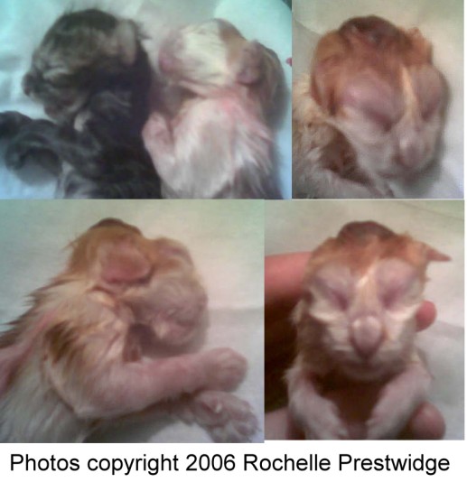
The image above was provided by Rochelle Prestwidge in October 2006. Rochelle's cat gave birth to 5 kittens of which 2 were abandoned by the mother. The second-born kitten (male) had anencephaly. Comparing the male to his sister (both were stillborn or died soon after birth), his head is flat from above the eyes straight across with a bulging red membranous section in the middle. Although closed, his eyes are noticeably larger and his lower jaw protrudes slightly. In comparison, the normal kitten's head is domed above the eyes.
Anencephaly is a neural tube defect that occurs when the cephalic (head) end of the neural tube fails to close early in gestation, resulting in the absence of a major portion of the brain, skull, and scalp. Affected individuals are born without a forebrain i.e. the largest part of the brain comprising mainly of the cerebrum which is responsible for thinking and coordination. Lack of cerebrum explains the kittens inability to co-ordinate and suckle. The remaining brain tissue is often exposed (not covered by bone or skin) and matches Joen's description of a visible membrane. The braincase does not form properly and this distorts other features, in this case the lidless prominent eyes and the placement of the ears. According to human medicine, anencephalic newborns are usually blind, deaf, unconscious and unable to feel pain. Some individuals have a rudimentary brainstem, but the lack of a functioning cerebrum makes consciousness impossible. Reflex actions such as breathing and responses to sound or touch may occur. In humans, the condition is found more often in females than in males. Affected individuals are usually stillborn, those that survive birth die within hours or, at most, a few days.
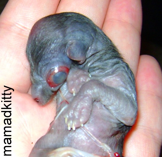
Another photo of a neural tube defect from "mamadkitty". Her cat, Paris had a litter of 3 kittens. The first Kitten was stillborn and appears to have anencephaly. The other 2 kittens, a male and a female, were normal. The photo was taken straight after birth and there has been no shrinkage of tissues.
This stillborn kitten was born 5 days premature and delivered placenta first. The hairlessness, bloating and the darkening under the skin indicates it had died some days earlier; this may have triggered the premature delivery of the litter. It appers to have died, or stopped developing physically, at a stage before the genitalia had differentiated into male or female.
MORE ANOMALIES
If you have come to this page directly from a search engine, please check out
FELINE MEDICAL CURIOSITIES for the full index of topics includingBOOKS ABOUT ANOMALIES
If you are interested in medical curiosities, books worth reading are "Mutants: on the Form, Varieties and Errors of the Human Body" by Armand Marie Leroi and "Anomalies and Curiosities of Medicine Vols 1 and 2" by George M. Gould & Walter L. Pyle. The Gould & Pyle books were published in 1896 and are in the public domain. You can download text-only versions of Gould & Pyle from several websites so don't waste money on text-only versions of the book; but if you want the versions with photos, consider the Kessinger editions. The Leroi book explains why and how some deformities and anomalies happen - the mechanism is the same in cats as it is in humans.
You are visitor number